
Nuclear Thyroid Imaging is one among the various functional and molecular imaging procedures being undertaken at Imaging Lily, with the help of much improved version of latest Nuclear Imaging equipments. The imaging services offered here are unique, as they are carried out conforming to the National regulations and International standards.
DIY IMAGING LILY, located in Padivattom near pipeline junction opposite to POC,on civil line road towards KAKANAD, is the first ever stand alone Pvt.Ltd, PET-CT centre in ‘‘The Spice Garden Of India’’, KERALA. It is committed to provide the best possible for the Health Care need of the Nation; in the realm of Nuclear Medicine and Radiology. Nuclear Thyroid Imaging is one among the various functional and molecular imaging procedures being undertaken here, with the help of much improved version of latest Nuclear Imaging equipments. The imaging services offered here are unique, as they are carried out conforming to the National regulations and International standards.

The thyroid gland is a two lobed, butterfly shaped organ located in front of the neck. It secretes thyroid hormones such as T3 (Triiodothyronine) and T4 (Tetraiodothyronine - Thyroxin).The levels of these hormones in our blood is controlled by pituitary gland through feedback mechanism by releasing Thyroid Stimulating Hormone(TSH).This hormone stimulates the thyroid gland either to increase or decrease the secretion of thyroid hormone according to the need. Thyroid hormones are very important for our body because they regulate the metabolic activities that are vital for the development of our brain, body and sexual organs.
Depending up on the status of the thyroid function, there are many classification of thyroid disease.
1. Functional abnormalities:
a) Hyperthyroidism - increased secretion of thyroid hormone.
b) Hypothyroidism - decreased secretion of thyroid hormone.
2. Nodular abnormalities:
a) Endemic Goitre.
b) Diffuse Goitre.
c) MNG-Multi Nodular Goitre.
3. Tumor:
a) Thyroid adenoma
b) Thyroid cancer:
i) Papillary
ii) Follicular
iii) Medullary
iv) Anaplastic.
4. Lymphomas and Metastases.
5. Deficiency: Congenital hypothyroidism-Cretinism.
6. Ectopic Thyroid:
a) Lingual Thyroid
b) Thyroglossal cyst.
Though biochemical tests and ultrasound imaging are available for diagnosing thyroid abnormalities, Nuclear Thyroid Scan gives a visual picture of the thyroid gland and its iodine trapping mechanism.
Nuclear Thyroid Scan is done to evaluate the functional status of thyroid gland. This is variously known as isotope thyroid scan, thyroid scintigraphy, etc. This scan is carried out by introducing a calculated quantity of a specific radionuclide in to our body. The distribution of radionuclide in the thyroid gland is mapped by a special equipment called Gamma Camera. Variation in the brightness of the image gives the variation of function in different regions of the thyroid gland. The image also gives the changes in the size, shape and location of the gland. Based on the image pattern and the uptake values, the different types of thyroid diseases are identified.
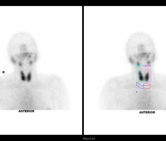
Fig.1
EUTHYROID STATUS.
Normal Thyroid Scan with uniform distribution of tracer and normal uptake value.
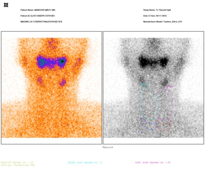
Fig.2
THYROIDITIS.
Normal Thyroid Scan with uniform distribution of tracer and normal uptake value.

Fig.3
HYPERTHYROIDISM.
Intense uniform distribution of tracer with high uptake. High T3, T4 values and low TSH level

Fig.4
HYPERTHYROIDISM WITH GOITRE :
Intense non uniform distribution of tracer with high T3 and T4 and low TSH values.

Fig.5
EUTHYROIDISM WITH NODULARITY :
MNG Non uniform tracer distribution with multi nodularity and normal uptake values.T 3 ,T 4 and TSH levels are normal.

Fig.6
HYPOTHYROIDISM WITH AGENESIS OF RIGHT LOBE:
Absence of right lobe with Non - uniform distribution of tracer in the left lobe. Low T 3 , T 4 and high TSH levels.

Fig.7 Autonomous nodule-TOXIC GOITRE

Fig.8 HYPER THYROIDISM WITH COLD NODULE

Fig.9 LINGUAL THYROID

Fig.10 THYROGLOSSAL CYST